Micro-computed tomography for structure-function investigation
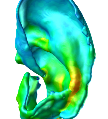
The ICRG approach uses micro-computed tomography with novel contrast agents (31), other imaging modalities (4, 9), imaging protocols (16) and processing methods to capture detailed structural information of organs, tissues and cells, and then links this to materials analyses; via mechanical testing and assays, to provide unique insight into the structure-function relationships. For example, in joints (5, 7, 25), ears (21), and tissue-engineered constructs (15).
Publications
31. B. A. Lakin, H. Patel, C. Holland, J. D. Freedman, J. S. Shelofsky, B. D. Snyder, M. W. Grinstaff and K. S. Stok. Contrast-enhanced CT using a cationic contrast agent enables non-destructive assessment of the biochemical and biomechanical properties of mouse tibial plateau cartilage, J. Orthop Res, 34(7):1130-1138, 2016.
25. C. Barnabe, S. Finzel, K. S. Stok, P. Geusens, Invited correspondence “High-resolution peripheral quantitative CT in rheumatology”, Nature Rev Rheum, 11(2):123, 2015.
21. L. Nimeskern, E. M. Feldmann, S. Schwarz, W. Kuo, S. Duerr, E. Goldberg-Bockhorn, R. Müller, N. Rotter, K. S. Stok, Magnetic Resonance Imaging of the ear for patient-specific reconstructive surgery, PLoS One, 9(8): e104975, 2014.
16. R. J. Choo, R. Müller and K. S. Stok. Prevention of cartilage dehydration in imaging studies with a customised humidity chamber. Rev. Sci. Instrum., 84(9), 093703, https://dx.doi.org/10.1063/1.4820913, 2013.
15. W. Kuo, L. Nimeskern, H. Martínez Ávila, S. Hofmann, J. Freedman, M. W. Grinstaff, R. Müller, K. S. Stok. Developing staining protocols for visualization of tissue-engineering scaffolds using micro computed tomography in native wet state. Proc. Gemeinsame Jahrestagung der Deutschen, österreichischen und Schweizerischen Gesellschaft für Biomedizinische Technik, Graz, Austria, September 19-21, Biomed. Tech., 58(S1):4273, 2013.
9. J. P. Moodie, K. S. Stok, R. Müller, T. L. Vincent and S. J. Shefelbine. Multimodal imaging demonstrates concomitant changes in bone and cartilage after destabilisation of the medial meniscus and increased joint laxity. Osteoarthr Cartilage, 19:163-170, 2011.
7. K. S. Stok, D. Noël, F. Apparailly, D. Gould, Y. Chernajovsky, C. Jorgensen and R. Müller. Quantitative imaging of cartilage and bone for functional assessment of gene therapy approaches in experimental arthritis. J. Tissue Eng. Regen. Med., 4:387-394, 2010.
5. K. S. Stok, G. Pelled, Y. Zilberman, I. Kallai, D. Gazit, and R. Müller. Revealing the interplay of bone and cartilage in osteoarthritis through multimodal imaging of murine joints, Bone, 45:414–422, 2009.
4. K. S. Stok, and R. Müller. Morphometric characterisation of murine articular cartilage – Novel application of confocal laser scanning microscopy, Microsc. Res. Techniq., 72:650–658, 2009.